


Sold out
About this collection
Medical Models for Anatomy Learning & Demonstration
Medical models provide a three-dimensional way to study and explain human anatomy. They can help medical students, physiotherapy students, teachers and healthcare professionals understand structures that may be harder to visualise from a textbook alone.
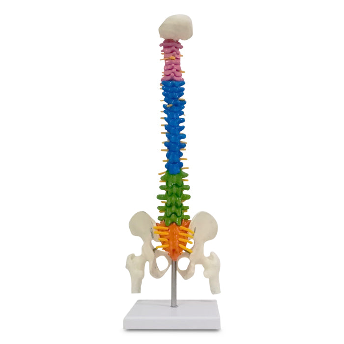
Meddey's current Bone Models collection features a compact colour-coded spine model. This model shows the main regions of the human spine in a format suited to classroom teaching, clinical demonstrations and patient education.
Unlike diagnostic equipment, anatomical models do not measure or treat a condition. Instead, they work as visual learning and communication aids. Buyers should therefore choose a model according to the anatomical structure they need to demonstrate.
Current Medical Model Available at Meddey
Mini Coloured Spine Model – 45 cm Tall
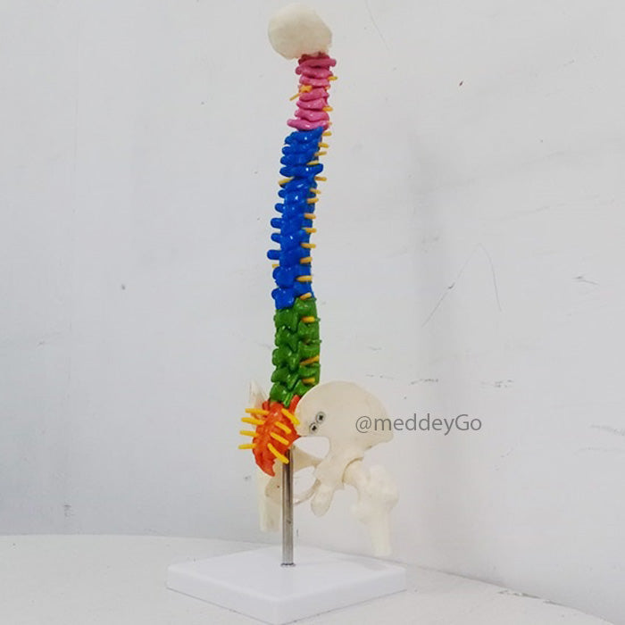
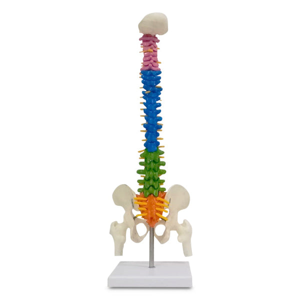
This compact spine model displays the major sections of the vertebral column in different colours. As a result, students and clinicians can identify each spinal region more quickly during demonstrations.
The model includes cervical, thoracic and lumbar vertebrae together with the sacrum and coccyx. Its 45 cm height makes it compact enough for a teaching desk, consultation room or classroom demonstration.
Colour-Coded Anatomy Learning
Colour differentiation helps users distinguish one spinal region from another. For example, students can visually separate the cervical area from the thoracic and lumbar sections without relying only on written labels.
Patient explanations may also become easier with a physical model. A healthcare professional can point directly to a spinal section while discussing basic anatomy or the location relevant to a consultation.
What Does the Spine Model Show?
The human vertebral column contains several distinct anatomical regions. On the current Meddey model, these areas use different colours to support quick visual identification.
- 7 cervical vertebrae: the vertebrae located in the neck region.
- 12 thoracic vertebrae: the mid-back region where the ribs connect.
- 5 lumbar vertebrae: the lower-back section of the spine.
- Sacrum: the triangular region located below the lumbar spine.
- Coccyx: the lower end of the vertebral column.
Since each region is visually separated, the model can support demonstrations of basic spinal organisation without requiring learners to identify every region from shape alone.
Who Can Use Medical Models?
Anatomical models have several educational uses. Medical and allied-health students can combine them with textbooks, diagrams and practical teaching sessions.
Physiotherapists may use a spine model when explaining basic spinal anatomy to patients. Meanwhile, anatomy teachers can demonstrate the relationship between different vertebral sections during lectures or practical classes.
Clinics can also keep compact medical models in consultation rooms for patient education. However, these models should remain educational aids rather than diagnostic tools.
Suitable Users
- Medical students
- Physiotherapy students
- Nursing and allied-health students
- Anatomy teachers and educators
- Physiotherapists
- Doctors and clinicians
- Medical colleges and training institutes
- Clinics using visual patient education
Why Use Medical Models for Anatomy Study?
Two-dimensional diagrams are useful for learning names and locations. Nevertheless, a physical model gives learners another way to understand the shape and arrangement of anatomical structures.
Viewing the vertebral column from different angles can reveal relationships that are less obvious in a flat illustration. Consequently, learners may find it easier to understand how the spinal regions connect.
Group teaching can also benefit from physical medical models. An instructor can point to the relevant structure while students observe the same model.
With careful handling, reusable anatomy models can support repeated demonstrations across different classes or consultations.
Medical Models for Students
Medical models can help students connect written anatomy with a physical representation of the body. A spine model supports this process by making vertebral regions easier to locate visually.
During revision, colour changes can act as useful reference points. Students may first identify the cervical, thoracic and lumbar regions and then compare them with textbooks, diagrams or other study material.
Compact dimensions also make the current model practical for a study table or teaching room. At 45 cm tall, it provides a visible representation without needing the space of a full-size skeleton model.
Medical Models for Clinics & Patient Education
In clinics, medical models can make spinal anatomy easier to explain. Verbal descriptions may be difficult for some patients to follow, so a physical model gives clinicians a clear visual reference.
A clinician might point to the cervical region when discussing the neck or to the lumbar region when describing lower-back anatomy. Even so, the model supports communication rather than replacing clinical assessment, medical imaging or professional diagnosis.
Colour-coded sections can make explanations easier to follow as well. Instead of relying only on anatomical terminology, patients can visually identify the region being discussed.
Medical Models for Classroom Teaching
Anatomy teachers can use a physical spine model while explaining the organisation of the vertebral column. Because the major regions use different colours, students have a clear visual reference during the lesson.
Teachers can also combine the model with diagrams projected on a screen. For instance, an instructor may introduce a vertebral region on a diagram before demonstrating its position on the three-dimensional model.
Smaller practical sessions may allow learners to examine the model more closely. Appropriate classroom handling helps protect it during repeated use.
How to Choose an Anatomy Model
When comparing medical models, start with the anatomical structure that needs to be studied. A spine model suits lessons focused on the vertebral column, whereas a skull or full skeleton model serves a different educational purpose.
Size should be considered next. Compact models work well for individual study, desk demonstrations and smaller classrooms, while larger models may suit situations where visibility from a distance matters more.
Level of detail is equally important. Colour coding can help learners distinguish different regions, whereas other model types may use labels, detachable parts or movable joints.
Storage requirements should also influence the final choice. A teaching aid used regularly needs a suitable location where it remains protected from unnecessary impact, dust and heat.
Mini Spine Model vs Full-Size Skeleton
| Feature | Mini Spine Model | Full-Size Skeleton Model |
|---|---|---|
| Main focus | Spinal anatomy | Whole-body skeletal anatomy |
| Space required | Compact | Considerably more space |
| Best suited to | Spine teaching, desk demonstrations and patient education | Whole-body anatomy teaching |
| Portability | Easier to move and store | Usually less portable |
| Current Meddey Bone Models range | Available | Not currently shown in this specific collection |
Understanding the Main Sections of the Spine
The cervical spine forms the neck portion of the vertebral column. It contains seven vertebrae and sits above the thoracic region.
Below the neck, the thoracic spine contains twelve vertebrae. This section forms the mid-back and connects with the ribs.
Five vertebrae make up the lumbar spine in the lower back. Moving further downward, the vertebral column continues into the sacrum and coccyx.
A colour-coded model makes this sequence easier to demonstrate because each major section can be identified visually during study or explanation.
Care & Storage of Anatomy Models
Regular care can help preserve an educational model for repeated use. Begin by removing surface dust with a soft, dry or slightly damp cloth when appropriate.
Harsh chemicals should be avoided unless the product instructions specifically allow them. Strong cleaning agents may affect painted or colour-coded surfaces.
A dry location away from excessive heat and direct sunlight is generally suitable for storage. Place the model where it is also unlikely to fall or receive unnecessary impact.
During practical sessions, students should handle the model carefully. Proper storage after each lesson can help preserve its appearance and structure over time.
Explore Related Meddey Collections
Students and educators looking for additional human-body teaching aids can explore Anatomy Models .
Rehabilitation professionals may also want to visit Rehabilitation Equipment for physiotherapy and recovery-related products.
People looking for braces and other supportive products can browse Orthopedic Supports .
Clinics that need examination and procedure tools can also explore Medical Instruments .
Frequently Asked Questions
What are medical models used for?
Medical models are mainly used for anatomy teaching, student learning, professional demonstrations and patient education. They provide a three-dimensional representation of anatomical structures.
What does the Mini Coloured Spine Model show?
The model shows the cervical, thoracic and lumbar vertebrae together with the sacrum and coccyx. Different colours separate the major spinal sections for easier identification.
How tall is the current spine model?
At 45 cm tall, the Mini Coloured Spine Model is suitable for desk, classroom and consultation-room demonstrations.
Is this medical model suitable for students?
Yes. Medical, physiotherapy, nursing and allied-health students can use the spine model as a visual aid alongside their normal anatomy study materials.
Can physiotherapists use a spine model?
Yes. Physiotherapists can use a spine model to demonstrate basic spinal anatomy and help patients identify the region being discussed during an appropriate consultation.
Can doctors use medical models for patient education?
Yes. Anatomical models can support explanations by giving patients a visual reference. They do not, however, replace examination, diagnosis or medical imaging.
Does this model include the lumbar spine?
Yes. Five lumbar vertebrae are shown along with the cervical and thoracic sections, sacrum and coccyx.
Is a mini spine model better than a full skeleton?
Each serves a different teaching need. A mini spine model focuses on the vertebral column and requires less space, while a full skeleton supports broader whole-body skeletal study.
How should an anatomy model be cleaned?
Gently remove dust and follow the product's care instructions. Avoid harsh chemicals that may damage coloured or painted surfaces.
Need Help Choosing Medical Models?
For current availability, product specifications or assistance with anatomy teaching models, contact Meddey Customer Care on WhatsApp at +91 85860 12345 .
Educational note: Anatomical models are teaching and demonstration aids. They do not provide diagnosis, treatment or an alternative to professional clinical assessment.
Explore Medical Collections
All Categories
Abdominal & Maternity Support

Accessories

Accu-Chek Instant Strips: Use, Compatibility, Price & Blood Sugar Testing Guide
Accu-Chek Instant Strips: Uses, Features & Complete Guide

Accupressure Products for Pressure Point Therapy, Pain Relief & Wellness

accupuncture

Acupuncture Needles & Supplies

Advanced Physiotherapy Machines & Lumbar Physiotherapy

Airways

Ambulance

Anatomy Models
Anesthesia Machine

Anesthesia Products
Ankle Support, Braces & Walker Boots for Recovery

Ankle Weights & Exercise Weights for Home & Rehab